Tag Archives: retinal damage symptoms

Effective Retinal Detachment Treatments: Options and Outcomes

Effective Retinal Detachment Treatments: Options and Outcomes

Retinal detachment is a serious condition that requires timely diagnosis and appropriate treatment to prevent vision loss. Understanding the different types of retinal detachment, the diagnostic techniques available, and the treatment options can help patients make informed decisions about their care. In this article, we will explore the various aspects of retinal detachment and discuss the effective treatments available.

Understanding Retinal Detachment

The Anatomy of the Eye

The eye is a marvel of biological engineering, comprising intricate components that harmoniously work together to enable the sense of vision. Among these components, the retina stands out as a vital player in the visual process. Positioned at the posterior part of the eye, the retina acts as a light-sensitive layer that captures incoming light rays and converts them into neural signals, which are then relayed to the brain for interpretation. This complex cascade of events is fundamental to our ability to perceive the world around us with clarity and detail.

Moreover, the retina is not a uniform structure but consists of distinct layers, each with specialized functions contributing to visual acuity. These layers work in concert to ensure that the visual information received by the eye is accurately processed and transmitted to the brain for meaningful interpretation. Any disruption to this intricate system, such as retinal detachment treatments, can have profound implications for vision and overall ocular health.

Effective Retinal Detachment Treatments: Options and Outcomes

Causes and Symptoms of Retinal Detachment

Retinal detachment can arise from a myriad of factors, ranging from sudden trauma to the eye, degenerative changes associated with aging, to underlying ocular conditions like myopia or diabetic retinopathy. The onset of retinal detachment is often heralded by a cluster of distinct symptoms that should not be ignored. These symptoms may manifest as the abrupt onset of floaters—small, dark spots drifting in the visual field, flashes of light akin to lightning bolts, and the perception of a shadow or curtain descending over the field of vision. Recognizing these warning signs and seeking immediate medical intervention is paramount in mitigating the risk of irreversible vision loss.

Furthermore, the treatment for retinal detachment typically involves surgical intervention aimed at reattaching the detached retina to its original position and restoring visual function. Various surgical techniques, such as scleral buckling or vitrectomy, may be employed based on the severity and extent of the detachment. Timely diagnosis and intervention play a pivotal role in the prognosis of retinal detachment, underscoring the importance of regular eye examinations and proactive eye care to safeguard ocular health.

Types of Retinal Detachment

Rhegmatogenous Retinal Detachment

Rhegmatogenous retinal detachment is the most common type and occurs when a tear or hole develops in the retina, allowing fluid to accumulate between the retina and its underlying tissue. This can lead to the detachment of the retina, impairing vision.

Rhegmatogenous retinal detachment often occurs spontaneously, but it can also be associated with trauma to the eye or previous eye surgeries. The accumulation of fluid behind the retina causes it to separate from the underlying layers, leading to symptoms such as sudden flashes of light, floaters in the field of vision, and a curtain-like shadow over the visual field. If left untreated, this type of detachment can result in permanent vision loss. To read more about vision loss click here.

Tractional Retinal Detachment

Tractional retinal detachment occurs when scar tissue or abnormal blood vessels exert traction on the retina, pulling it away from its normal position. Conditions such as diabetic retinopathy can contribute to this type of detachment.

In diabetic retinopathy, the growth of abnormal blood vessels can cause fibrous scar tissue to develop on the surface of the retina. As this scar tissue contracts, it can distort the shape of the retina and pull it away from the underlying tissues, leading to tractional retinal detachment. Patients with this condition may experience gradual blurring of vision, distortion of shapes, and difficulty seeing in low light conditions.

Exudative Retinal Detachment

Exudative retinal detachment occurs when fluid leaks into the area between the retina and its supportive tissues, usually due to a disorder affecting the blood vessels. This type of detachment is less common but can still cause vision problems.

Conditions such as age-related macular degeneration or inflammatory disorders can lead to exudative retinal detachment. In these cases, abnormal blood vessels or inflammation can cause fluid to accumulate under the retina, leading to a separation of the layers. Patients with exudative retinal detachment may experience a sudden decrease in central vision, distortion of straight lines, and difficulty recognizing faces.

Diagnosis of Retinal Detachment

Clinical Examination

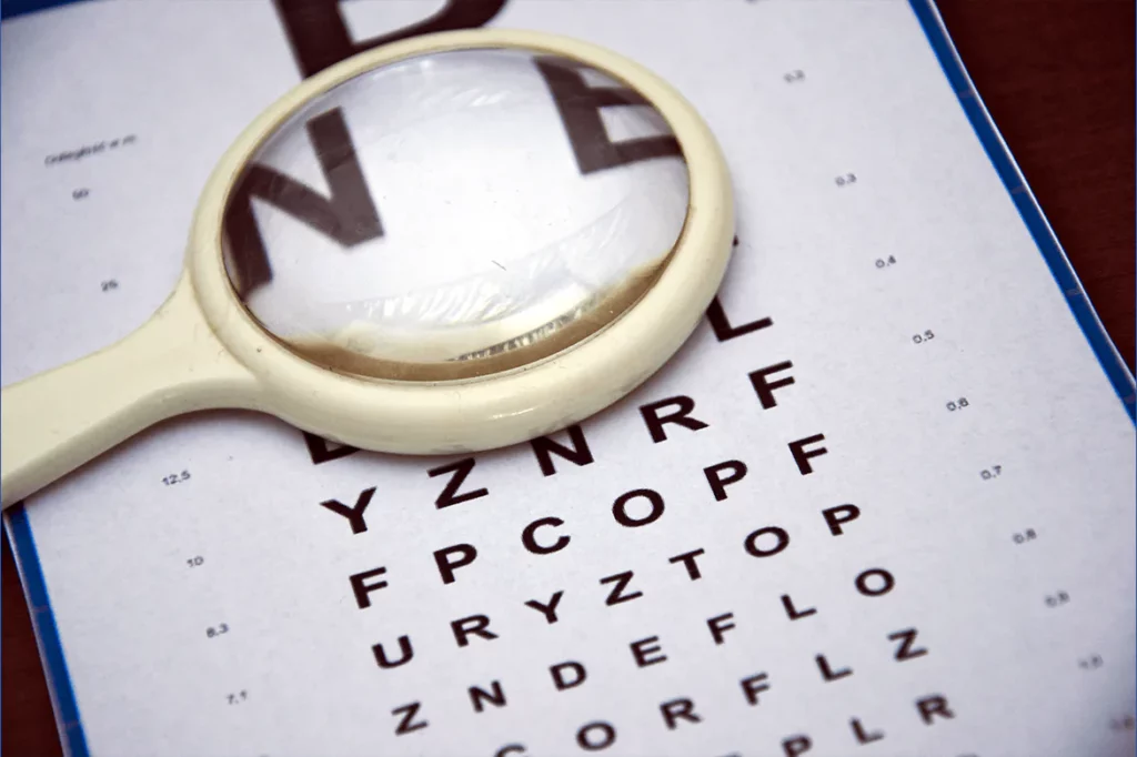
Diagnosing retinal detachment typically involves a comprehensive eye examination. Your ophthalmologist will evaluate your symptoms, conduct a visual acuity test, and examine the back of your eye using specialized instruments to assess the condition of the retina.

During the clinical examination, your ophthalmologist will also perform a detailed assessment of your medical history to identify any underlying conditions or risk factors that may contribute to retinal detachment. This holistic approach helps in determining the most appropriate treatment plan tailored to your individual needs.

Imaging Techniques

In addition to a clinical examination, your ophthalmologist may utilize imaging techniques such as optical coherence tomography (OCT) or ultrasound to gather more detailed information about the retina and determine the extent and location of the detachment.

Optical coherence tomography (OCT) is a non-invasive imaging test that uses light waves to capture high-resolution cross-sectional images of the retina. This technology allows for the visualization of the different layers of the retina, helping the ophthalmologist to precisely locate any areas of detachment and monitor the response to treatment over time.

Treatment Options for Retinal Detachment
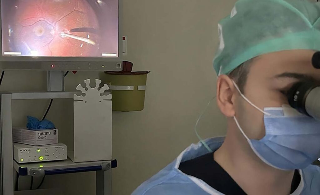
Surgical Procedures

Surgery is the most common method for treating retinal detachment. Depending on the type and severity of the detachment, different surgical techniques may be employed. These procedures aim to reposition the retina and seal any tears or holes to prevent further detachment.

One common surgical procedure for retinal detachment is scleral buckling. During this procedure, a silicone band is placed around the eye to gently push the wall of the eye against the detached retina, helping it to reattach. Another surgical option is vitrectomy, where the vitreous gel inside the eye is removed and replaced with a gas bubble to push the retina back into place. Learn more about vitrectomy at https://www.urmc.rochester.edu/encyclopedia/content.aspx?contenttypeid=135&contentid=385

Laser Therapy

Laser therapy, also known as photocoagulation, uses focused beams of light to create small burns around the retinal break or tear. This process helps to create scar tissue that seals the retina and prevents further detachment.

It is important to note that laser therapy is often used for small retinal tears or holes that have not yet progressed to a full detachment. The procedure is typically quick and can be done in an outpatient setting, with minimal discomfort for the patient. However, laser therapy may not be suitable for all types of retinal detachments, and your ophthalmologist will determine the most appropriate treatment plan for your specific condition.

Cryotherapy

Cryotherapy involves the use of extreme cold to freeze the area around the retinal tear or detachment. Freezing the tissue creates scars, which act as barriers to prevent further separation of the retina.

While cryotherapy can be effective in treating retinal detachments, it is often used in combination with other surgical techniques for optimal results. The procedure is performed under local or general anesthesia, and the cold temperature is carefully controlled to ensure the safety of the surrounding eye structures. Cryotherapy may cause some temporary discomfort or redness in the eye, but these symptoms typically resolve as the eye heals.

Effective Retinal Detachment Treatments: Options and Outcomes

Post-Treatment Care and Recovery

Follow-up Visits and Monitoring

Following retinal detachment treatment, close monitoring is essential to ensure proper healing and detect any recurrent or new changes. Regular follow-up visits with your ophthalmologist will allow them to evaluate your progress and make any necessary adjustments to your treatment plan.

During these follow-up visits, your ophthalmologist will conduct thorough examinations of your eyes, which may include visual acuity tests, intraocular pressure measurements, and detailed retinal evaluations. These evaluations help in assessing the success of the treatment and identifying any signs of complications early on. Your ophthalmologist will also discuss any symptoms or concerns you may have, providing you with the opportunity to address them promptly.

Lifestyle Adjustments for Recovery

During the recovery phase, it is important to take certain precautions to optimize healing. Your ophthalmologist may recommend avoiding activities that put strain on the eyes, such as heavy lifting or vigorous exercise. Additionally, proper nutrition and maintaining a healthy lifestyle can facilitate recovery.

Engaging in relaxation techniques, such as deep breathing exercises or meditation, can also be beneficial during the recovery period. These practices not only help reduce stress and anxiety but also promote overall well-being, which is essential for the healing process. Adequate rest and sleep are equally important, as they allow your body, including your eyes, to recuperate and regenerate effectively.

Read more

Recognizing Retinal Damage Symptoms: Early Signs and Prevention

Recognizing Retinal Damage Symptoms: Early Signs and Prevention

Retinal damage is a condition that affects the retina, a vital part of the eye responsible for transmitting visual information to the brain. This condition can cause vision loss and potentially lead to blindness if left untreated. Recognizing the early signs of retinal damage is crucial for timely intervention and prevention of further deterioration. In this article, we will explore the various aspects of retinal damage, including its causes, symptoms, the importance of early detection, prevention strategies, and treatment options.

Understanding Retinal Damage

The retina is a complex and vital component of the eye, playing a crucial role in our ability to see. This thin layer of tissue is composed of several layers of cells, each with a specific function in the process of vision. The photoreceptors, including rods and cones, are responsible for capturing light and converting it into electrical signals. These signals are then processed by other retinal cells before being transmitted to the brain via the optic nerve.

Retinal Damage Symptoms is a condition that can have a significant impact on vision if left untreated. Recognizing the early signs of retinal damage and seeking prompt medical attention is crucial for preventing further deterioration. Regular eye examinations, adopting a healthy lifestyle, and considering preventive measures can also contribute to maintaining retinal health.

Furthermore, the health of the retina is closely linked to the overall health of the body. The retina receives its oxygen and nutrients from a network of blood vessels known as the retinal vasculature. Any disruption to this blood supply can have detrimental effects on the retina, leading to potential damage and vision loss.

What is Retinal Damage?

Retinal damage encompasses a wide range of conditions that can impact the structure and function of the retina. This damage can manifest in various forms, from minor changes in vision to severe vision impairment or even blindness. It is essential to address retinal damage promptly to prevent further deterioration and preserve vision.

Recognizing Retinal Damage Symptoms: Early Signs and Prevention

Causes of Retinal Damage

While aging is a common factor in retinal damage, other systemic conditions can also contribute to its development. Hypertension and diabetes, for example, can affect the blood vessels in the retina, leading to conditions such as diabetic retinopathy or hypertensive retinopathy. These conditions highlight the intricate connection between systemic health and ocular health, emphasizing the importance of comprehensive healthcare and regular eye examinations.

In addition to systemic conditions, trauma to the eye can also result in retinal damage. Blunt force trauma, penetrating injuries, or even excessive exposure to harmful light can all have detrimental effects on the delicate structures of the retina. Understanding the potential causes of retinal damage is crucial in implementing preventive measures and seeking timely treatment to preserve vision and overall eye health.

Identifying Early Symptoms of Retinal Damage

Early detection of retinal damage symptoms is essential for timely intervention and management. Being aware of these symptoms can help individuals seek appropriate medical attention and prevent the progression of the condition.

Retinal damage refers to any injury or deterioration that affects the retina, the light-sensitive tissue lining the back of the eye. The retina plays a crucial role in converting light into neural signals that are sent to the brain for visual recognition. Damage to the retina can result in various visual disturbances and, if left untreated, may lead to permanent vision loss. Read more about vision loss at https://hms.harvard.edu/news/vision-revision

Common Symptoms of Retinal Damage

Symptoms of retinal damage can vary depending on the underlying cause and the specific area of the retina affected. Some common symptoms include:

  • Blurred or distorted vision
  • Reduced color perception
  • Floaters, which are dark spots or specks that appear to float in the field of vision
  • Flashes of light
  • Difficulty seeing at night

If you experience any of these symptoms, it is important to consult an eye care professional for a comprehensive examination.

Unusual Symptoms You Shouldn’t Ignore

While the above-mentioned symptoms are commonly associated with retinal damage, there are also some unusual symptoms that should not be ignored. These include sudden, painless vision loss, the appearance of a curtain-like shadow over the visual field, or a sudden increase in the number of floaters. These symptoms could indicate a more severe condition and require immediate medical attention.

Additionally, individuals with retinal damage may experience a gradual loss of peripheral vision or a sensation of seeing “wavy” or crooked lines. In some cases, patients may also report a sudden onset of double vision or a significant decrease in visual acuity. These symptoms can be indicative of underlying retinal issues that need to be addressed promptly to prevent further damage.

The Importance of Early Detection

Early detection of retinal damage can significantly impact the outcome and treatment options available. By identifying the condition at an early stage, healthcare professionals can implement appropriate interventions to prevent further damage and preserve vision.

Retinal damage, if left untreated, can lead to irreversible vision loss and even blindness. The retina is a crucial part of the eye responsible for capturing light and converting it into neural signals that are sent to the brain for visual recognition. Damage to the retina can disrupt this process, resulting in blurry vision, blind spots, or total loss of sight. Therefore, early detection is key to maintaining optimal eye health and preserving vision for as long as possible.

How Early Detection Can Prevent Further Damage

When retinal damage is detected early, treatment options such as medication, laser therapy, or surgery can be initiated promptly. These interventions aim to stabilize the condition, slow down the progression of damage, and prevent vision loss. Early detection also allows for the implementation of lifestyle modifications and preventive measures to maintain retinal health.

Furthermore, early intervention can improve the effectiveness of treatment and increase the likelihood of successful outcomes. For conditions like age-related macular degeneration or diabetic retinopathy, early detection and timely treatment can help preserve central vision and prevent severe complications that may result in permanent vision impairment.

The Role of Regular Eye Examinations

Regular eye examinations play a crucial role in the early detection of retinal damage. Eye care professionals can conduct comprehensive evaluations, including visual acuity tests, dilated eye exams, and imaging studies to assess the condition of the retina. It is recommended to schedule regular eye check-ups, especially for individuals at higher risk of retinal damage, such as those with a family history of retinal diseases or underlying health conditions like diabetes.

During an eye examination, the optometrist or ophthalmologist may also evaluate other aspects of eye health, such as intraocular pressure, optic nerve health, and overall eye function. These comprehensive assessments not only aid in the early detection of retinal issues but also help in the timely diagnosis and management of other eye conditions, ensuring overall visual well-being.

Prevention Strategies for Retinal Damage

While certain risk factors for retinal damage, such as age and genetics, cannot be controlled, there are proactive steps individuals can take to promote retinal health and reduce the risk of damage.

Retinal damage is a serious concern that can lead to vision impairment or even blindness if left untreated. The retina is a thin layer of tissue located at the back of the eye that is responsible for converting light into neural signals, which are then sent to the brain for visual recognition. Damage to the retina can occur due to various factors, including age-related macular degeneration, diabetic retinopathy, retinal detachment, and other eye conditions.

Lifestyle Changes for Retinal Health

Adopting a healthy lifestyle can have a positive impact on retinal health. This includes maintaining a balanced diet rich in antioxidants and nutrients, regular exercise, managing chronic conditions like diabetes and hypertension, avoiding smoking, and protecting the eyes from prolonged exposure to harmful UV rays by wearing sunglasses. To know more about prolonged exposure to harmful UV rays click here.

Foods rich in antioxidants, such as leafy greens, fish, nuts, and citrus fruits, can help protect the retina from oxidative stress and inflammation. Regular physical activity promotes overall health, including proper blood circulation to the eyes, which is essential for maintaining retinal function. Managing conditions like diabetes and hypertension is crucial, as uncontrolled high blood sugar and blood pressure levels can damage the blood vessels in the retina, leading to vision problems.

Medical Interventions for Prevention

In some cases, medical interventions may be recommended to prevent or slow down the progression of retinal damage. These interventions can include medications, such as anti-VEGF drugs, which help reduce abnormal blood vessel growth in the retina, or laser therapy to seal leaking blood vessels. Early detection and consultation with a healthcare professional can guide the appropriate preventive options based on individual circumstances.

Regular eye examinations are essential for detecting early signs of retinal damage or other eye conditions. An ophthalmologist can perform specialized tests, such as optical coherence tomography (OCT) or fluorescein angiography, to assess the health of the retina and identify any abnormalities. By monitoring the retina regularly, potential issues can be addressed promptly, potentially preventing irreversible damage and preserving vision.

Recognizing Retinal Damage Symptoms: Early Signs and Prevention

Navigating Treatment Options

When it comes to treating retinal damage, various options are available depending on the severity and underlying cause. Treatment plans are personalized to the individual’s condition and may involve a combination of non-surgical and surgical interventions.

Non-Surgical Treatments for Retinal Damage

Non-surgical treatments for retinal damage include medications, such as corticosteroids or anti-inflammatory drugs, which can help reduce inflammation and edema in the retina. These medications work by suppressing the immune response and reducing the release of inflammatory substances in the eye. By doing so, they can alleviate symptoms and slow down the progression of retinal damage.

Additionally, intraocular injections of anti-VEGF drugs can be administered to inhibit abnormal blood vessel growth and manage conditions like age-related macular degeneration (AMD) or diabetic retinopathy. These drugs target vascular endothelial growth factor (VEGF), a protein that promotes the growth of abnormal blood vessels in the retina. By blocking the action of VEGF, these injections can help prevent further damage to the retina and preserve vision.

Surgical Treatments for Advanced Cases

In advanced cases of retinal damage, surgical interventions may be necessary. Procedures like retinal detachment repair, vitrectomy, or macular hole surgery aim to restore or stabilize the retina’s structure and function. Retinal detachment repair involves reattaching the detached retina to the underlying tissue, while vitrectomy involves removing the vitreous gel from the eye and replacing it with a clear solution. Macular hole surgery, on the other hand, focuses on repairing a hole in the macula, the central part of the retina responsible for sharp, detailed vision.

These surgeries are typically performed by ophthalmologists specialized in retinal conditions. They require precision and expertise to ensure the best possible outcome for the patient. The surgeon will carefully assess the individual’s condition, determine the most appropriate surgical approach, and discuss the potential risks and benefits of the procedure.

With advancements in medical interventions, timely treatment options are available to manage retinal damage and preserve vision. However, it is important to remember that every case is unique, and treatment plans should be tailored to the individual’s specific needs. If you experience any symptoms or have concerns about your retinal health, consult an eye care professional for a comprehensive evaluation. They will be able to provide you with the necessary guidance and recommend the most suitable treatment options for your condition.

Read more.